Differentiating Monophasic Synovial Cell Sarcoma of the Kidney from Other Tumors of Kidney and Review of the Literature
Tanushri Mukherjee
DOI10.21767/2471-299X.1000035
Tanushri Mukherjee*
Oncopathologist, Command Hospital, Kolkata, India
- *Corresponding Author:
- Tanushri Mukherjee
Oncopathologist, Associate Professor
Command Hospital, Kolkata, India
Tel: 8697989701
E-mail: tanujamukherjee@yahoo.com
Received date: September 15, 2016; Accepted date: September 25, 2016; Published date: September 30, 2016
Citation: Mukherjee T. Differentiating Monophasic Synovial Cell Sarcoma of the Kidney from Other Tumors of Kidney and Review of the Literature. Med Clin Rev. 2016, 2:3.doi: 10.21767/2471-299X.1000035
Copyright: © 2016 Mukherjee T. This is an open-access article distributed under the terms of the Creative Commons Attribution License, which permits unrestricted use, distribution, and reproduction in any medium, provided the original author and source are credited.
Keywords
Monophasic synovial sarcoma; Spindle cell tumor; bcl2
Background
Synovial sarcoma is commonly a tumor of soft tissue which is seen in the extremities, abdominal wall, neck, head, mediastinum and abdominal cavity. Synovial sarcoma may be biphasic or monophasic. Monophasic Synovial cell sarcoma (SCS) of the kidney is a rare tumor first described by Faria et al. and documented in 2000 by Argani et al. [1]. Only few cases are reported in literature [2,3]. It accounts for 5–10% of adult soft tissue sarcomas. There is no clinical or imaging characteristic that can indicate the diagnosis. The diagnosis is difficult due to rarity of tumor and its similar presentations as compared to other renal tumors [4,5]. It mostly affects young people and is associated with bad prognosis and is a rare tumor in location of kidney and so causing diagnostic difficulty and differentiating it from other renal malignant tumors is important which are common like sarcomatoid renal cell carcinoma, sarcomas and hemangiopericytoma Monophasic synovial sarcoma is composed of spindle cells only and there is no epithelial component and biphasic is having spindle cells with admixed epithelial cell component and there may be poorly differentiated type of synovial sarcoma. Immunohistochemistry is characteristic with CD99, bcl2 and TLE11 and there is SYT-SSX gene fusion rearrangement p11.2 and q11.2 of chromosomes X and 18, respectively, i.e., t (X; 18) (p11.2; q11.2) [4]. We report: A case of a young man who underwent right radical nephrectomy at our institution.
Case Report
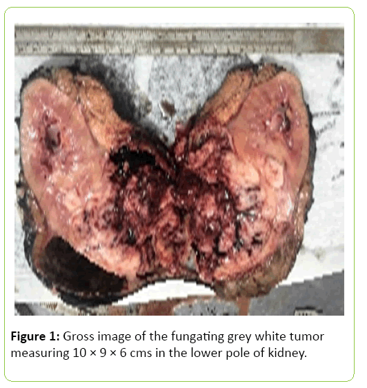
32 years old male patient reported with right kidney mass and loin pain. The patient was a non-smoker and had no history of exposure to any chemicals. Right radical nephrectomy was performed. Right renal vein showed a thrombus. The gross of the kidney showed weight 320 g and measuring 15 × 12 × 10 cms with a fungating grey white tumor measuring 10 × 9 × 6 cms in the lower pole involving medulla and pelvis and renal capsule, perinephric fat and gerotas fascia (Figure 1).
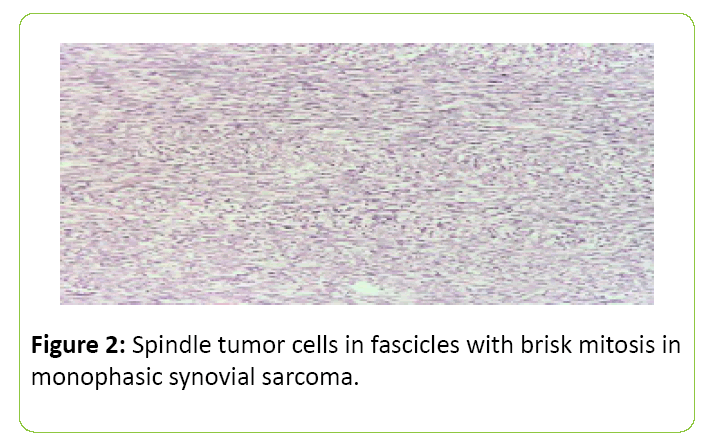
Microscopy showed a monotonous tumor composed of only spindle-shaped tumor cells. There was prominent mitotic activity and a microscopic renal vein vascular invasion (Figure 2).
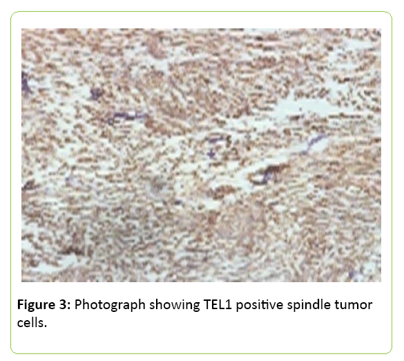
Immunohistochemical analysis demonstrated positivity for Bcl-2, CD99, transducin-like enhancer of split 1 (TLE1) and vimentin (Figure 3). The other markers such as CK, S100, HMB45, SMA, actin, CD34, desmin, CK22 and CK19 was negative. There was no epithelial component and because of characteristic immunohistochemistry it was called as monophasic SCS. Diagnosis confirmed by SYT gene rearrangement by FISH. The patient was kept under regular follow up.
Discussion
Monophasic synovial sarcoma of the kidney is extremely rare tumor and less than 50 cases have been described in the literature. This tumor affects predominantly males and young patients. There are no tell-tale specific clinical or radiological features of this tumor in kidney so diagnosis is extremely difficult and is very important to differentiate due to bad prognosis. Histologically monophasic synovial sarcomas may be difficult to differentiate this from sarcomatoid tumors originating in the kidney, especially sarcomatoid renal cell carcinoma, retroperitoneal sarcomas, malignant peripheral nerve sheath tumor, primitive neuroectodermal tumors involving the kidney [6-10]. Immunohistochemistry with TEL1, bcl2, CD99 and vimentin is helpful. Confirmation of diagnosis is by rearrangement involving the SYT gene by FISH or PCR. Radical surgical resection remains the treatment of choice. The use of adjuvant chemotherapy is recommended for younger patients and larger tumors where clinical advantages could rather be expected. The prognosis is poor with this treatment of surgery alone. Synovial Sarcoma may be sensitive to high dose isophosphamide and adriamycin based regimen. As the number of cases of Synovial Sarcoma of the kidney is less due to its extreme rarity, no clear medical guidelines have yet been established [11]. Accurate diagnosis is important as these tumors known to behave aggressively and bad outcome with recurrence and metastases [12].
Conclusion
The incidence of monophasic synovial sarcoma of the kidney is less and due to its extreme rarity, no clear medical guidelines have yet been established and accurate diagnosis is important as these tumors known to behave aggressively and bad outcome with recurrence and metastases.
References
- Argani P, Faria PA, Epstein JI, Reuter VE, Perlman EJ, et al. (2000) Primary renal synovial sarcoma: Molecular and morphologic delineation of an entity previously included among embryonal sarcomas of the kidney. Am J SurgPathol 24:1087-1096.
- Mauricio A, Paláu L, Pham TT, Barnard N, Merino MJ (2007) Primary synovial sarcoma of the kidney with rhabdoid features. Int J SurgPathol 15:421-428.

Open Access Journals
- Aquaculture & Veterinary Science
- Chemistry & Chemical Sciences
- Clinical Sciences
- Engineering
- General Science
- Genetics & Molecular Biology
- Health Care & Nursing
- Immunology & Microbiology
- Materials Science
- Mathematics & Physics
- Medical Sciences
- Neurology & Psychiatry
- Oncology & Cancer Science
- Pharmaceutical Sciences


