Gastric Adenocarcinoma with Mixed Histology in a 29-Year-Old Patient
Zelaya KAT, Maradiaga JJM, Pastor AT and Avila LHI
DOI10.21767/2471-299X.1000030
Zelaya KAT1,2*, Maradiaga JJM3,4, Pastor AT5 and Avila LHI5
1Faculty of Medical Sciences, National Autonomous University of Honduras, Tegucigalpa, Honduras
2Medical Science University of Havana Cuba, Minimal Access Surgery Center, Hondurus
3Faculty of Medical Sciences, National Autonomous University of Honduras, Tegucigalpa, Honduras
4Universidad de San Carlos de Guatemala, Roosevelt Hospital, Guatemala
5Faculty of Medical Sciences, Catholic University of Honduras, Campus Sacred Heart of Jesus, Comayagüela, Honduras
- *Corresponding Author:
- Zelaya KAT
Faculty of Medical Sciences
National Autonomous University of Honduras
Tegucigalpa, Honduras and Medical Science University of Havana Cuba
Minimal Access Surgery Center, Hondurus
Tel: +50494621010/+50427851408
E-mail: arielat_29@hotmail.com
Received date: August 17, 2016; Accepted date: August 25, 2016; Published date: September 02, 2016
Citation: Zelaya CAT, Maradiaga JJM, Pastor AT, et al. Gastric Adenocarcinoma with Mixed Histology in a 29-Year-Old Patient. Med Clin Rev. 2016, 2:3. DOI: 10.21767/2471-299X.1000030
Copyright: ©2016 Zelaya CAT, et al. This is an open-access article distributed under the terms of the Creative Commons Attribution License, which permits unrestricted use, distribution, and reproduction in any medium, provided the original author and source are credited.
Abstract
The gastric carcinoma is one of the three primary malignant gastric neoplasms. Sorted by Lauren as, the intestinal type adenocarcinoma (53%) which is frequent in elderly and males (2:1), the diffuse type gastric adenocarcinoma (33%) with a higher incidence in women between 30 and 40 years old; they are voluminous, aggressive and have a poor prognosis. Finally, the poorly differentiated or mixed gastric adenocarcinoma (14%) which is frequent in elderly and male patients. We present the case of a 29-year-old female patient from the city of Catacamas, Olancho, Honduras. Initially attended by a general practitioner with symptoms of epigastric burning pain treated as gastritis. Later on, the symptoms were exacerbated and accompanied by gastroesophageal reflux, gastric fullness, weight loss and sometimes dark feces; according to the anamnesis, the symptoms where presented by at least 6 months. The fact of having no history of malignancy in the family is included in the clinic history. Weeks later, she was examined by an internist in the same city, who referred her to “Clínica y Hospital Tróchez Montalván” in Juticalpa, Olancho, Honduras for upper tract endoscopy. The upper tract endoscopy revealed: diffuse, friable and stony tumor lesion covering a portion of the gastric body and the whole pylorus with thickening in its walls. Histopathology confirmed gastric adenocarcinoma of intestinal type, poorly ulcerated, focal areas with signet ring cells and infestation by fungal structures, Candida albicans compatible. The enzymatic and histological tests for Helicobacter pylori was reported negative. The patient relatives decided to take her for treatment to the Hospital Emma Romero de Callejas with chemotherapy. At this hospital she received one dose of chemotherapy and palliative care. The patient died one month later after being diagnosed. It is due to late diagnosis of such an aggressive disease as gastric cancer that brings awareness to people who have gastrointestinal symptoms to look for healthcare and alert health practitioners to diagnose and treat promptly, avoiding its development to a metaplastic aggression.
Keywords
Gastric Adenocarcinoma; Stomach; Cancer; Young Patient
Introduction
Gastric cancer is the second leading cause of death worldwide associated with cancer. Its incidence depends on the region being analyzed [1-5]. Gastric cancer is a disease that affects more the elderly and males (male: female; 2:1). In black people it is twice as common. Sometimes gastric cancer may present in young adults (diffuse infiltration) more often in women (3:1) [1]. Early diagnosis of gastric cancer is difficult, because it rarely presents with any symptoms in its initial stages. The most frequent symptoms and signs include: weight loss, loss of appetite, abdominal pain, vague abdominal discomfort (supraumbilical), epigastric pain, dyspepsia, satiety and fullness after eating a small meal, nausea, vomiting with or without blood, anemia, ascites, dysphagia, steatorrhea, belching etc. [6-11]. The diagnostic study of choice is gastric endoscopy (gastroduodenoscopy) and biopsy, with a sensitivity of 70% depending on the skill of the endoscopist. In 95% of gastric cancers they are classified as a gastric adenocarcinoma. After histopathological diagnosis, tests should be performed. The definitive treatment is surgery which depends on the tumor stage [1,6,9,11-13].
Honduras is a poor and under-developed country in Central America. According to clinical experience there has been an increase in the diagnosis of gastric cancer. Most of the studies and treatments are to be paid by patients, which is a limitation for proper management [14].
Clinical Case
Female patient, 29 years old from the city of Catacamas, Olancho, Honduras. Initially staffed by general practitioner with symptoms of epigastric pain. She was treated as gastritis. Later on, the symptoms were exacerbated and accompanied by gastroesophageal reflux, gastric fullness, weight loss and sometimes dark feces; according to the anamnesis, the symptoms where presented by at least 6 months. The fact of having no history of malignancy in the family was included. Weeks later, she was examined by an internist in the same city, who referred her to the “Clinica y Hospital Tróchez Montalván” in Juticalpa, Olancho, Honduras. She was received on May 6, 2016 in the Endoscopy Unit. Patient was stable, feverless, asthenic, and pale. Vital signs where: BP: 90/60 mmHg, HR: 63 beats per minute, Temperature: 37.2 degrees Celsius. Laboratory studies show: Hb 8.2 g/dL, Htc: 27% Leukocyte count: 7300 cells/mm3 Neutrophil count: 66% and Platelets: within normal range 232,000 cells/mm3.
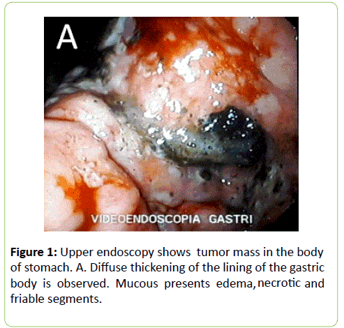
Upper gastrointestinal tract endoscopy was performed, noticing a malignant-looking ulcerated lesion infiltrating the gastric body and antrum. Multiple biopsies were taken from the mass for additional studies. Receiving biopsy results on May 11, 2016 which reveals:
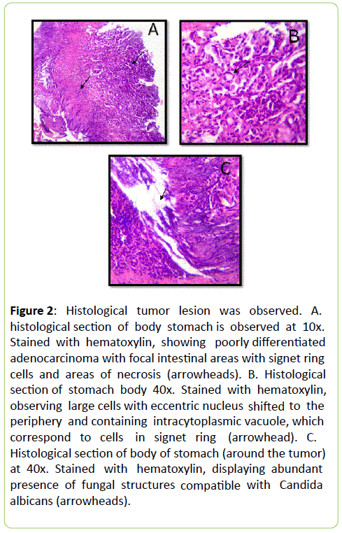
• Intestinal Adenocarcinoma infiltrative type poorly differentiated with abundant necrosis, old hemorrhage and recent mixed inflammatory infiltrate.
• Areas with signet ring cells.
• Fungal infestation structures compatible with Candida albicans.
• Negative Helicobacter pylori test. This study was conducted in Laboratory of Pathology of Olancho (Pathology and Cytology) by a pathologist.
Making an analysis of the results of the biopsy the presence of intestinal and diffuse adenocarcinoma was found in a single mass. Approximately two weeks after receiving positive results for gastric cancer the patient relatives decide to take her for treatment to the Emma Romero de Callejas Hospital where she started treatment with chemotherapy and palliative care. The patient died one month later after the diagnosis, June 10th of 2016.
Discussion
Gastric cancer is the second leading cause of death worldwide associated with cancer. Its incidence depends on the region being analyzed. Gastric cancer is more common in men than in women (2:1). The three most common primary malignant gastric neoplasms are Adenocarcinoma (95%), Lymphoma (4%) and Gastrointestinal Stromal Malignant Tumor (1%) [1]. The gastric adenocarcinoma according to the Lauren and Javi classification is divided into: 1) Intestinal (53%); 2) Diffuse (Signet Cell Ring) (33%); 3) Not classified/ mixed (14%). Intestinal adenocarcinoma corresponds to a well differentiated gastric carcinoma. It’s characterized by an expansive growth pattern and resembles intestinal glands and epithelial cells. Diffuse type adenocarcinoma (undifferentiated) corresponds to the infiltrative variety. They contain dispersed cells that invade individually and may observe signet cell ring and mucin. This extends a few millimeters from primary tumor [1-5,10,14-17]. Intestinal Adenocarcinoma is more common in the elderly and males in 53%. The adenocarcinoma has an incidence of 33% in women 30-40 years old who have a poor prognosis because of its size and aggressiveness [1-2].
Early diagnosis of gastric cancer is difficult because it rarely presents symptoms in the initial stage. The most frequent symptoms and signs include weight loss, loss of appetite, abdominal pain, vague abdominal discomfort (supraumbilical), epigastric pain, dyspepsia, satiety and fullness after eating a small meal, nausea, vomiting with or without blood, anemia, ascites, dysphagia, steatorrhea, belching etc. [6-12]. The finding of gastric cancer in young patients depends on clinical suspicion by the physician. The gastroduodenoscopy is the best diagnostic method. This method allows visualization of the mass and facility to obtain a sample for histological study. The greater number of biopsies taken increase the possibility of obtaining a more accurate result. One biopsy has an accuracy of 70%, four biopsies of 95% and rises to 98% with 7 or more biopsies. If the tumor mass is exophytic, endoscopy generally allows for a tissue diagnosis. We can also perform diagnostic studies such as esophagus-gastro-duodenal endoscopic, ultrasound and computed tomography(Figure 1) [8,10-12,17-19].
The etiology of gastric cancer is multifactorial; however, the most outstanding is the Helicobacter pylori in 60% of the cases. Helicobacter pylori causes continuous inflammation of the gastric mucous. When Helicobacter pylori invades gastric mucous there is a recruitment of neutrophils, T and B lymphocytes, plasma cells, macrophages, IL-8, tumor necrosis factor α, increased p53 and interferon. Which leads to decreased antral cells causing hypochlorhydria. Helicobacter pylori infection induces the formation of mucosa-associated lymphoid tissue (MALT) in the gastric tissue. Causing gastriculcer (s) if not eradicated. Gastric ulcer and atrophic gastritis are main precursors of gastric cancer. Helicobacter pylori causes gastritis and may produce gastric atrophy, in some patients it leads to intestinal metaplasia and later on to dysplasia [1,3,4,20-23].
Risk factors are multiple. It has been observed that gastric cancer is more common in patients with pernicious anemia, blood type A or family history. Its incidence according to gender is more common in men than in women. Most gastric cancer is diagnosed in patients between 60-89 years of age. Rich carbohydrate and nitrite diet raises the risk of a possible gastric cancer [1,6,9-10,19].
Because cancer patients are generally considered immunosuppressed they are at high risk of opportunistic infections such as Candida albicans. Candida albicans has the capacity to grow and multiply in the bowels and may have access to the bloodstream producing fungal planting especially in the liver by the portal circulation. Candida albicans infection increases the mortality rate by 39% and this percentage increases if the patient is elder (Figure 2) [24].
Figure 2: Histological tumor lesion was observed. A. histological section of body stomach is observed at 10x. Stained with hematoxylin, showing poorly differentiated adenocarcinoma with focal intestinal areas with signet ring cells and areas of necrosis (arrowheads). B. Histological section of stomach body 40x. Stained with hematoxylin, observing large cells with eccentric nucleus shifted to the periphery and containing intracytoplasmic vacuole, which correspond to cells in signet ring (arrowhead). C. Histological section of body of stomach (around the tumor) at 40x. Stained with hematoxylin, displaying abundant presence of fungal structures compatib l e with Candida albicans (arrowheads).
References
- Brunicardi FC, Andersen DK,Billiar TR, Dunn DL, Hunter JG (2006) Schwartz Principles of Surgery (Eighth ed.) Mexico: McGraw Hill Interamericana, 2006.
- Anquela S (1989) Importance of Lauren classification of gastric cancer. Revision of a series of 295 cases. Club de Patologia Digital Sociedad Española de anatomiaPatologica. 22:156-161.
- Martinez-Galindo MG (2014) Histopathologic features of gastric adenocarcimoa in mesxicanos patient. Experiencia de 10 años en el Hospiyal Juarez de Mexico. Revista de Gastroenterologia de Mexico 1:21-26.
- Lopez-BasaveHN (2013) Gastric cancer in young people under 30 years of age: worst prognosis, or delay in diagnosis? Cancer Manag Res 5:31-36.
- Pisanu A, Podda M, Cois A, Uccheddu A (2014) Gastric Cancer in the Young: Is it a Different Clinical Entity? A Restrospective Cohort Study. Gastroenterology Research and Practice9.
- Society AC (2016) Stomach cancer. American Cancer Society.
- Rivas DJA (1982) Gastric Cancer in Honduras. RevistaMedica de Honduras 5:68-70.
- Turcios-Galeano RE, Vargas-Pineda C (1998) Epidermiology of Gastric Cancer in Honduras: The Experience in the Hospital Escuela. Rev Med Post Unah Mayo a Agosto 3:100-112
- Arana Reyes JC, Bautista AC (2004) Gastric cancer. Revista de la Facultad de Medicina UNAM 47: 204-209.
- AdeLTB (2010) Guide diagnosis and treatment of gastric cancer. Diagnosis. J Gastroenterol Mexico.
- Seker M (2013) Clinicopathologic features of Gastric Cancer in Young Patients. Saudi J Gastroenterol 19:258-261.
- Subsecretaria de SaludPublica (2014) Gastric cancer. GuiaClinica. Chile: Plan de acceso Universal de GarantiasExplicitas, Ministerio de Salud.
- Dominguez RL,Crockett SD, Lund JL, Suazo LP, Heidt P, et al. (2013) Gastric Cancer Incidence Estimation in a Resource Limited Nation: Use of Endoscopy Registry Metholodology. Cancer Causes Control.
- Wang Y (2014)Clinicpathological Features of Gastric Cancer in Young Patients. J Cancer Ther 5:1366-1371.
- Gómez M, Otero W, Caminos JE (2012) Gastric cancer in young patients in Colombia. Rev Col Gastroenterol27:166-172.
- Romero LHE, Siancas JN (2003) Classification of stomach adenocarcinomas. Revista de estrienterologia de Peru 23:199-212.
- Cañon JS (2003) Cell carcinoma of the stomach signet ring knowledge of its biology or natural history. ActaCancerologica 32:3-4.
- Park HJ (2014) Clinical Characteristics and Outcomes for Gastric Cancer Patients Aged 18-30 years Old. Gastric Cancer: The International Gastric Association and The Japanese Gastric Cancer Association 17:649-660.
- Isobe T, Hashimoto K, Kizaki J, Miyagi M, Aoyagi K(2013) Characteristics and prognosis of gastric cancer in young patients. Oncol Rep 30: 43-49.
- gil-NegreteA (2013) Protocols for Gastric Cancer Management. Protocolo de Actuacion. España: Osakidetza-euskadi, ONCOLOGIA; Report No.: ss-396/2014.
- Dr. Posse RSA, Dr. Toledo RA, Dr. Cabral MLV (2006) Helicobacter Pylori: Clinic, Diagnosis and Treatment. Revistas de Posgrado de VIaCatedra de Medicina 158:9-12.
- Veauchamp EM (2013)SabistonTratado de Cirugia(19thedn.) Barcelona, España: Elsevier; Townsend.
- Dhobi MA (2013) Gastric Cancer in Young Patients. International Journal of Surgical Oncology4.
- Garcia-Ruiz JC, Amutio E, Pontón J (2004) Invasive fungal infection in immunocompromised patients. RevistaIberoamericanaMicologia21:55-62.

Open Access Journals
- Aquaculture & Veterinary Science
- Chemistry & Chemical Sciences
- Clinical Sciences
- Engineering
- General Science
- Genetics & Molecular Biology
- Health Care & Nursing
- Immunology & Microbiology
- Materials Science
- Mathematics & Physics
- Medical Sciences
- Neurology & Psychiatry
- Oncology & Cancer Science
- Pharmaceutical Sciences


