Implications of CBCT in Dentistry: A Review
Aakash Shah
DOI10.21767/2471-299X.1000057
Aakash Shah*
Department of Orthodontics and Dentofacial Orthopedics, K.M. Shah Dental College and Hospital, Sumandeep Vidyapeeth University, India
- *Corresponding Author:
- Shah A
BDS, MDS, Department of Orthodontics and Dentofacial Orthopedics
K.M. Shah Dental College and Hospital
Sumandeep Vidyapeeth University, India
Tel: + 91-9825656377; +91-8200894584
E-mail: aakashshah108@gmail.com
Received date: September 27, 2017; Accepted date: October 13, 2017; Published date: October 23, 2017
Citation: Shah A (2017) Implications of CBCT in Dentistry- A Review. Med Clin Rev. 3:15. doi: 10.21767/2471-299X.1000057
Copyright: © 2017 Shah A. This is an open-access article distributed under the terms of the Creative Commons Attribution License, which permits unrestricted use, distribution, and reproduction in any medium, provided the original author and source are credited.
Abstract
Cone beam Computed Tomography (CBCT) is acclaimed for its accuracy and diverse clinical utility. The benefits of good image quality, volumetric analysis, short scan times, and relatively less radiation dose than conventional medical CT, has resulted in greater ubiquity as an imaging modality within all disciplines of dentistry. It has become an important adjunct in orthodontic diagnosis due in part to the diverse image reconstructions available (cephalometrics, TMJ cross-sections, etc.), the ability to visualize bony levels, and the sub-millimeter accuracy enabling linear measurements. In particular, evaluating fine anatomical structures, like alveolar bone enveloping teeth, is important to the orthodontist for both initial diagnostic knowledge and outcome assessment. The ability to characterize duccal bone has clear benefits for practitioners in periodontics and implant dentistry as well.
Keywords
CBCT; Craniofacial imaging
Introduction
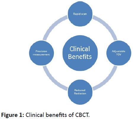
Cone beam computed tomography (CBCT) is an absolutely recent imaging technology used to create 3-dimensional renditions of subjects [1]. Following the commercial introduction of CBCT, unprecedented abilities to maxillofacial imaging emerged, immensely expanding the role of imaging within diagnostics and treatment [1]. The benefits of good image quality, volumetric analysis, short scan times, and relatively less radiation dose than conventional medical CT, has resulted in greater ubiquity as an imaging modality within all disciplines of dentistry. Many fields, including orthodontics, oral surgery, implant dentistry, periodontics, and endodontics find unique utility of the 3-dimensional reconstructions provided by CBCT [2,3]. It has become an important adjunct in orthodontic diagnosis due in part to the diverse image reconstructions available (cephalometrics, TMJ cross-sections, etc.), the ability to visualize bony levels, and the sub-millimeter accuracy enabling linear measurements [4,5].
Shortcomings of CBCT
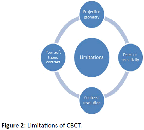
However, CBCT is known to have shortcomings, such as capturing thin areas of bone [6-8]. The accurate imaging of these fine anatomical structures is important to the orthodontic clinician for both initial diagnostic decisions and outcome assessment as the radiographic interpretation of bone levels is often used to determine periodontal health or externalities as the result of treatment [2,3,9].
Components of CBCT
There are many components of CBCT image production; the various factors, such as the scanning unit employed, examined object, FOV, contrast resolution, and spatial resolution defined by the voxel size may profoundly influence the image quality produced for interpretation [10]. It is important to understand these details in order to pursue improvement in the modality on a clinical level. The immediately following paragraphs will elaborate on this detail to establish the foundation for discussion of the scientific study of these variables, and how manipulation will produce different clinical results.
Imaging from the CBCT is accomplished via a rotating gantry, from which a pyramidal x-ray beam is directed through the subject onto a contralateral sensor [1]. The gantry will rotate around the subject simultaneously collecting multiple (from 150 to more than 600), sequential, full-volume, planar projection (2D) images within an assigned field of view (FOV), each individually known as basis images [1]. These basis images are used to mathematically reconstruct the 3- dimensional volume for viewing and manipulation [1].
With the collection of each individual image in CBCT geometry, the full volume of the subject is scanned, generating a significant amount of omnidirectional scatter that is ultimately recorded by the receptor [1]. This reduces image contrast and increases image noise [5]. The larger the area, or field of view, of the scan, the more scatter generated. The fields of view imaged by CBCT are adjustable and collimation of the x-ray beam limits exposure to the region of interest, allowing the operator to narrow the scope of the image for each individual patient and clinical need [1]. Naturally, the larger fields of view are associated with larger amounts of exposure [11].
Contrast resolution refers to the ability of an observer to distinguish between two objects of different radiographic densities [12]. High contrast between the margins of an object and surrounding structures improves the observer ability to identify those boundaries at the interface [5,12]. Therefore, the narrow interface between tooth structure and the enveloping bone, with similar radiodensities, would be more difficult to distinguish than that between air and bone [5,12].
Spatial resolution is the minimum distance necessary to distinguish between two objects [5]. With CBCT-derived images, spatial resolution, and therefore detail, is primarily defined by individual volume elements or, voxels [1]. A voxel is the 3D equivalent to the 2D pixel, whereby a voxel is defined by its height, width, and depth [10]. CBCT voxels are generally isotropic, meaning equal in all dimensions [10]. The area detector resolution of CBCT units is sub-millimeter, ranging from 0.09 to 0.40 mm, principally determining the size of the voxels [1]. Reducing the voxel size during a scan will improve the resolution, at a cost of increased radiation exposure to the patient [5,13]. Thus, the voxel dimension utilized is directly related to the radiation dose to which the patient is submitted during the scan [7].
Clinical benefits
Effective radiation dose to the patient (ranging from 29-477 μSv) relative to traditional medical CT (approximately 2000 μSv) is greatly reduced (1,14). Multiple, interactive display renditions developed for unique diagnostic and operative clinical needs allow prodigious clinical flexibility [1].
Limitations
A considerably large amount of factors are integral to the production of CBCT volumes [5]. This intricate and multifactorial nature of CBCT image production raises many questions. Operationally adjustable parameters such as FOV and spatial resolution (voxel size) change the diagnostic outcome of CBCT generated images [10]. However, the consequences, the magnitude of those consequences, and the appropriate clinical applications are all poorly understood. For medical CT examinations, settings or protocols for any application are well established [10]. Conversely, rationales for standardized protocols and their impact on CBCT-based diagnosis are presently unavailable for dentistry [10].
This is important for the clinician to judiciously utilize CBCT technology, adhering to the ALARA principle of maximizing clinical benefit to the patient and minimizing the risks inherent to ionizing radiation [11,15,16].
Various studies
Many studies exist, employing a diverse variety of methodologies that demonstrate the ability of CBCT to produce accurate images. Initial reporting on accuracy began in 2004, with two notable publications by Kobayashi et al. and Lascala et al. [17,18]. Kobayashi used cadaver mandibles and Lascala used dry skulls, each comparing actual measurements to those made on CBCT, concluding that it is reliable for linear measurements of structures closely associated with dentomaxillofacial imaging [17,18].
Studies began to manipulate scanning parameters such as FOV and voxel size, in order to evaluate the outcome on image quality. In early 2010, Damstra et al. used dry mandibles embedded with glass spheres to evaluate the linear accuracy of CBCT generated surface models with 2 different voxel sizes (0.40mm and 0.25mm) and concluded accuracy in the CBCT measurement procedure with no significant difference between the voxel resolutions [19]. An inherent, and acknowledged, limitation in this study was the lack of soft tissues, resulting in increased contrast of the landmarks, influencing the outcome [19].
With demonstrable accuracy over long distances, investigation in the limits of spatial resolution emerged. Sun et al. used pig specimens to measure alveolar bone height from CBCT generated images with varying voxel sizes [12]. These authors found evidence that decreasing voxel size improved the accuracy of alveolar bone measurements [12]. In an effort to most closely emulate a clinical setting, Patcas et al. used intact cadaver heads and evaluated the ability of CBCT, with varying voxel resolutions, to detect the bony covering of mandibular anterior teeth [6]. Differing in conclusions by Damstra regarding the significance of voxel resolutions, Patcas results, along with the earlier report by Sun et al. [12] suggest demonstrated improvement in accuracy when decreasing the voxel size [6]. Despite that improvement however, the authors went on to discuss that differences between clinical and radiographic measurements can be as large as 2mm, showing that the average alveolar bone thickness of 1mm might be missed completely [6]. Overall, this report concluded that CBCT is an appropriate tool for linear measurements and that the presence of surrounding tissue as well as different voxel size affects the precision of the data [6]. It was acknowledged that even the most granular 0.125-mm voxel protocol does not depict the thin buccal alveolar bone covering reliably, resulting in a risk of overestimating fenestrations and dehiscences [6]. This unreliability is substantiated by Leung et al., who found that CBCT has a high rate of false positives with 3 times the number of fenestrations detected than existed in reality and a significant number of false negatives with more than half of real dehiscences undetected [20].
In an article recently published by Cook et al, the authors varied scanning parameters and measured buccal alveolar bone height and thickness on human cadavers. Their protocol compared images generated from a “long scan” with 619 basis images, 360° revolution, 26.9s duration, and 0.2mm voxel size against those from a “short scan” with 169 basis images, 180 rotation, 4.8s duration, and 0.3mm voxel size; the measurements made from these scans were compared to direct caliper measurement [21]. The authors found no statistically significant differences between these parameter changes and concluded that the parameters resulting in a lower radiation dose to the patient was favorable unless the need for the higher resolution could be clearly defined [21].
Studies with variations in voxel size during analysis extend beyond the evaluation of bone landmarks and measurement. A systematic review conducted by Spin-Neto et al. collated 20 different publications which qualitatively or quantitatively assessed the influence of voxel size on CBCT-based diagnostic outcome [10]. The diagnostic tasks evaluated in the studies included in the review were diverse, including detection of root fractures, detection of external root resorption, caries detection, and accuracy bony measurements, among others [10]. Some of the included studies demonstrated improvement in image quality and diagnostic accuracy, while others presented no difference [10,19,22,23-27]. Aggregately, the studies dealing with categorical data showed a tendency towards more accurate results associated with higher voxel resolutions [10]. However, Spin-Neto concluded that it is not yet possible to propose general protocols for the myriad of diagnostic applications with CBCT [10]. With the lack of unanimity, all of these investigations emphasize the need for a better understanding of the factors that influence image resolution with current CBCT technology.
Many authors have further discussed a concept referred to as the partial volume averaging effect [1,5,10,12]. This effect is a cone beam related artifact in which, depending on the voxel size, radiopaque structures could become invisible. As defined by Scarfe and Farman, partial volume averaging occurs when the selected voxel resolution of the scan is greater than the spatial or contrast resolution of the object to be imaged [1]. Meaning, the voxel is larger than the anatomical structure imaged and captures the image of two objects of different radiodensities. This voxel will then render the average density of both objects rather than the true density of either object [10]. Selection of the smallest acquisition voxel can reduce the effect of this averaging [1]. Known limitations in contrast resolution associated with CBCT units could also contribute to the invisibility of structures with similar radiodensities in close proximity [10,12]. The deficiencies as a result of low contrast resolution and partial volume averaging acknowledged by many authors are important to understand, and are critical concepts in future CBCT research.
Although CBCT has a relatively lower radiation dose to patients than medical CT, practitioners must be prudent in prescribing imaging in adherence to the ALARA principle (radiation dose ‘as low as reasonably achievable’). A myriad of factors contribute to the radiation exposure, among which are the aforementioned user adjustable settings of voxel size and FOV [1,11]. Other factors include scan duration, milliamperage, kilovolt potential, filtering, patient positioning, and the sensor technology and proprietary algorithms used in the device itself [1]. All of this makes CBCT dosimetry inherently difficult to summarize [11]. To further obfuscate, much of the research available relies on different methodologies and comparisons to draw conclusions about radiation exposure. DeVoss et al. conducted a systematic review of CBCT in 2009 and discussed findings on radiation dose in the literature. These authors found inconsistencies in how CBCT device settings, properties, and radiation dose were reported; all contributing to reader confusion [28]. They stated the importance of rigorous and consistent reporting on the different relevant parameters and acquisition protocols since device settings, image quality, and the resulting radiation exposure are closely related [28]. In 2013, Rottke et al. studied the effective dose (ED) span of ten different commercially available CBCT devices [14]. Performing protocols with the lowest exposition parameters and protocols with the highest exposition protocols for each of the ten devices, they found a wide range of EDs [14]. The average value for the protocols with the lowest exposition parameters was 31.6 μSv and 209 μSv for protocols with the highest exposition parameters.
Conclusion
The fundamental principle of utilizing CBCT for diagnosis and treatment planning in dentistry is to maximize the clinical benefit for the patient while minimizing the risks of ionizing radiation. The CBCT modality offers diverse utility but should be used prudently with the relationship between dose and image quality carefully considered. With all of its diverse uses and technical variability, dentistry has yet to develop standardized CBCT examination protocols. The development of these protocols would help guide practitioners when prescribing this modality for the wide variety of clinical applications.
References
- Scarfe WC, Farman AG (2008) What is cone-beam CT and how does it work?. Dent Clin North Am 52: 707-730.
- Rungcharassaeng K, Caruso JM, Kan JY, Kim J, Taylor G (2007) Factors affecting buccal bone changes of maxillary posterior teeth after rapid maxillary expansion. Am J Orthod Dentofacial Orthop 132: e1-8.
- Roe P, Kan JYK, Rungcharassaeng K, Caruso JM, Zimmerman G, et al. (2012) Horizontal and vertical dimensional changes of peri-implant facial bone following immediate implant placement and provisionalization of maxillary anterior single implants: A 1- year cone beam computed tomography study. Int J Oral Maxillofac Implants 27: 393-400.
- Lamichane M, Anderson NK, Rigali PH, Seldin EB, Will LA (2009) Accuracy of reconstructed images from cone-beam computed tomography scans. Am J Orthod Dentofacial Orthop 136: e1-6.
- Molen AD (2010) Considerations in the use of cone-beam computed tomography for buccal bone measurements. Am J Orthod Dentofacial Orthop 137: 130-135.
- Patcas R, Muller L, Ullrich O, Peltomaki T (2012) Accuracy of cone-beam computed tomography at different resolutions assessed on the bony covering of the mandibular anterior teeth. Am J Orthod Dentofacial Orthop 141: 41-50.
- Menezes CC, Janson G, Massaro CS, Cambiaghi L, Garib DG (2010) Reproducibility of bone plate thickness measurements with Cone-Beam Computed Tomography using different image acquisition protocols. Dental Press J. Orthod 15: 143-149.
- Leung CC, Palomo L, Griffith R, Hans MG (2010) Accuracy and reliability of cone-beam computed tomography for measuring alveolar bone height and detecting bony dehiscences and fenestrations. Am J Orthod Dentofacial Orthop 137: 109-119.
- Wood R, Sun Z, Chaudhry J, Ching Tee B, Kim D, et al. (2013) Factors affecting the accuracy of buccal alveolar bone height measurements from cone-beam computed tomography images. Am J Orthod Dentofacial Orthop 143: 353-363.
- Spin-Neto R, Gotfredsen E, Wenzel A (2013) Impact of Voxel Size Variation on CBCT- Based Diagnostic Outcome in Dentistry: a Systematic Review. J Digit Imaging 26: 813-820.
- Pauwels R, Beinsberger J, Collaert B, Theodorakou C, Rogers J, et al. (2011) Effective doze range for dental cone beam computed tomography scanners. Eur J Radiol 81: 267-271.
- Sun Z, Smith T, Kortam S, Kim DG, Tee BC, et al. (2011) Effect of bone thickness on alveolar bone-height measurements from cone-beam computed tomography images. Am J Orthod Dentofacial Orthop 139: e117-127.
- Kwong JC, Palomo JM, Landers MA (2008) Image quality produced by different cone-beam computed tomography settings. Am J Orthod Dentofacial Orthop 133: 317-327.
- Rottke D, Patzelt S, Poxleitner P, Schulze D (2013) Effective dose span of ten different cone beam CT devices. Dentomaxillofac Radiol 42: 20120417.
- Ludlow JB, Walker C (2013) Assessment of phantom dosimetry and image quality of i-CAT FLX cone-beam computed tomography. Am J Orthod Dentofacial Orthop 144: 802-817.
- Ludlow JB, Ivanovic M (2008) Comparative dosimetry of dental CBCT devices and 64-slice CT for oral and maxillofacial radiology. Oral Surg Oral Med Oral Pathol Oral Radiol Endod 106: 106-114.
- Kobayashi K, Shimoda S, Nakagawa Y, Yamamoto A (2004) Accuracy in measurement of distance using limited cone-beam computerized tomography. Int J Oral Maxillofac Implants 19: 228-231.
- Lascala CA, Panella J, Marques MM (2004) Analysis of the accuracy of linear measurements obtained by cone beam computed tomography (CBCT-NewTom). Dentomaxillofac Radiol 33: 291-294.
- Damstra J, Fourie Z, Huddleston Slater JJ, Ren Y (2010) Accuracy of linear measurements from cone-beam computed tomography-derived surface models of different voxel sizes. Am J Orthod Dentofacial Orthop 137: e1-6.
- Leung CC, Palomo L, Griffith R, Hans MG (2010) Accuracy and reliability of cone-beam computed tomography for measuring alveolar bone height and detecting bony dehiscences and fenestrations. Am J Orthod Dentofacial Orthop 137: 109-119.
- Cook VC, Timock AM, Crowe JJ, Wang M, Covell Jr DA (2015) Accuracy of alveolar bone measurements from cone beam computed tomography acquired using varying settings. Orthod Craniofac Res 18: 127-136.
- Liedke GS, da Silveira HE, da Silveira HL, Dutra V, de Figueiredo JA (2009) Influence of voxel size in the diagnostic ability of cone beam tomography to evaluate simulated external root resorption. J Endod 35: 233-235.
- Kamburoglu K, Murat S, Yuksel SP, Cebeci AR, Paksoy CS (2010) Occlusal caries detection by using a cone-beam CT with different voxel resolutions and a digital intraoral sensor. Oral Surg Oral Med Oral Pathol Oral Radiol Endod 109: 63-69.
- Kamburoglu K, Kursun S (2010) A comparison of the diagnostic accuracy of CBCT images of different voxel resolutions used to detect simulated small internal resorption cavities. Int Endod J 43: 798-807.
- Dalili Z, Taramsari M, Mousavi Mehr SZ, Salamat F (2012) Diagnostic value of two modes of cone-beam computed tomography in evaluation of simulated external root resorption: an in vitro study. Imaging Sci Dent 42: 19-24.
- Bauman R, Scarfe W, Clark S, Morelli J, Scheetz J, et al. (2011) Ex vivo detection of mesiobuccal canals in maxillary molars using CBCT at four different isotropic voxel dimensions. Int Endod J 44: 752-758.
- Maret D, Telmon N, Peters OA, Lepage B, Treil J, et al. (2012) Effect of voxel size on accuracy of 3D reconstructions with cone beam CT. Dentomaxillofac Radiol 41: 649-655.
- De Vos W, Casselman J, Swennen GRJ (2009) Cone-beam computerized tomography (CBCT) imaging of the oral and maxillofacial region: A systematic review of the literature. Int J Oral Maxillofac Surg 38: 609-625.

Open Access Journals
- Aquaculture & Veterinary Science
- Chemistry & Chemical Sciences
- Clinical Sciences
- Engineering
- General Science
- Genetics & Molecular Biology
- Health Care & Nursing
- Immunology & Microbiology
- Materials Science
- Mathematics & Physics
- Medical Sciences
- Neurology & Psychiatry
- Oncology & Cancer Science
- Pharmaceutical Sciences


