Primary Synchronous Bilateral Mucosa-Associated Lymphoid Tissue Lymphoma (MALT) of the Breast: A case Report with Discussion of Management and Review of the Literature
Daniele Colombo, Alessandro Bombonati, Vincenzo Ciocca, Lawrence Solin
DOI10.21767/2471-299X.1000012
Fondazione PTV Policlinico Tor Vergata, Rome, Rome Italy
- *Corresponding Author:
- Daniele Colombo
Fondazione PTV Policlinico Tor Vergata, Rome, Rome Italy
Tel: 06-2090-1
E-mail: dan.colombo82@gmail.com
Received Date: November 25, 2015; Accepted Date: December 30, 2015; Published Date: January 04, 2016
Citation: Daniele Colombo,Alessandro B,Lawrence S,Primary Synchronous Bilateral Mucosa-Associated Lymphoid Tissue Lymphoma (MALT) of the Breast: A case Report with Discussion of Management and Review of the Literature.Med Clin Rev. 2015, 2:3. doi: 10.21767/2471-299X.1000012
Copyright: © 2016 Daniele Colombo, et al. This is an open-access article distributed under the terms of the Creative Commons Attribution License, which permits unrestricted use, distribution, and reproduction in any medium, provided the original author and source are credited.
Abstract
Mucosa associated lymphoid tissue (MALT) lymphoma is a lymphoid neoplasm arising in extranodal sites with histologic features similar to those of nodal marginal zone lymphoma. Among the sites where a primary MALT lymphoma arises, the GI tract is the most common whereas the breast represents only 4%. Bilateral involvement of the breasts by a primary MALT lymphoma is exceptional with only 10 cases reported in the literature up to date. Herein, a case of a 73 years old woman with bilateral breast involvement by MALT lymphoma is presented and discussed.
Introduction
Mucosa associated lymphoid tissue (MALT) lymphoma is a lymphoproliferative disease arising in extranodal sites with histologic features similar to those of nodal marginal zone lymphoma. The most common site of origin of a MALT lymphoma is the gastrointestinal tract with the stomach being the most frequently involved site [1]. MALT lymphoma can arise from native mucosa-associated lymphoid tissue, such as tonsils or ileum, or from MALT that has been acquired as a result of a chronic inflammatory input or an autoimmune disease [2]. Organs where MALT is not usually present are GI tract, Lung, Salivary glands, Ocular adnexa and Breast [3].
Case Report
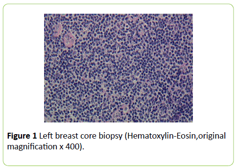
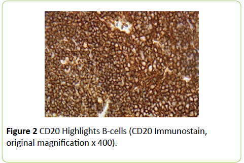
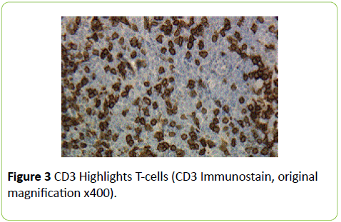
A 73 years old woman, with a history of uterine adenocarcinoma, hypertension and ocular myasthenia gravis, was found to have an indeterminate nodule in each breast (BIRADS- Breast Imaging-Reporting and Data System category 4) on bilateral mammograms and ultrasound. No other lesions or masses were noted in the rest of the body according to a pretreatment work-up consisting of Positron emission tomography-computed tomography (PET/CT) and blood work. Ultrasound-guided needle core biopsies of both right and left breasts showed a proliferation of atypical lymphocytes (Figure 1). Immunostains were initially performed on the left core biopsy (Figures 2 and 3) and the lymphocytic cell population was positive for CD20, CD79a (subset) and Bcl2, negative for CD10, CD23, Bcl6 and cyclinD1. CD21 was positive in follicular dendritic cells. CD138, kappa and lambda stains showed rare polytypic plasma cells. CD3 and CD5 highlighted background T cells. Histological and immunohistochemical features were highly suspicious for a low grade B cell neoplasm, favoring extranodal marginal zone lymphoma. Fluorescence in situ hybridization (FISH) analysis for Immunoglobulin heavy locus (IgH), which is commonly associated with lymphoid disorders, was performed and the result was negative for IgH gene rearrangement. Subsequently the patient underwent right and left breast lumpectomy. Although grossly the specimens did not show any discrete nodule or masses, the histological analysis showed an atypical lymphoid cell proliferation with features identical to those present in the previous core biopsies. The morphologic changes were consistent with the diagnosis of extranodal marginal zone B cell lymphoma. Additionally the neoplastic cells resulted positive for B-cell gene rearrangement by Polymerase chain reaction (PCR), further supporting the morphologic impression. The clinical staging bilaterally was I-EA. The patient received a course of definitive radiation treatment to the right breast and lower axilla and to the left breast and lower axilla, using high tangent fields bilaterally to a dose of 39.6 Gy. The patient tolerated the course of bilateral breast radiation treatment without complication. PET CT imaging and patient’s physical examination do not show any residual disease at the most recent follow up, 1 year and 2 months after treatment.
Discussion
Primary Breast lymphomas (PBLs) can be unilateral or bilateral [5]. It has been noted that bilateral breast lymphomas most commonly affect pregnant or lactating women. These lesions are characterized by an aggressive behavior [5], with their pathologic correlate being a Burkitt’s type lymphoma [6,7]. This aggressive behavior results in widespread dissemination to the ovaries and central nervous system [4]. PBLs represent less than 1% of all patients with NHL and account for between 0.04% and 1% of all breast malignancies [8]. Up to 50% of PBLs are Diffuse Large B Cell Lymphomas (DLBCL) [9].
Among the most common sites involved by MALT lymphoma, breast represents only 4% [10]. Martinelli et al. analyzed 264 cases of PBLs from 278 patients enrolled in the International Extranodal Lymphoma Study Group (IELSG). 204 (77%) cases were DLBCL, 36 (13%) were Follicular Lymphomas, while 24 (10%) resulted to be extranodal Marginal Zone Lymphomas (MZL aka MALT). Only 1 patient had a bilateral MZL [11]. 5 of the 24 patients with MZL were treated with radiation, 5 with surgery and 1 with chemotherapy. Surgery, in association with chemotherapy or radiation or both chemo and radiation, was performed on 13 patients with MZL-PBL. In this study, the majority of patients with MZL received radiotherapy as part of the initial treatment, which was usually delivered after surgery or biopsy. Most of the relapses occurred in distant sites, with only 21% of the recurrences arising in the primary disease sites (13%) or in the controlateral breast (8%). No patients who received radiotherapy relapsed within the irradiated fields, confirming the role of this modality treatment to prevent local recurrence. Bilateral breast involvement by MALT lymphoma is rare, with only ten cases reported in the literature up to date [11-21]. A review of the literature points out that the majority of patients, with early stage unilateral or bilateral MALT breast lymphoma, has been treated with radiotherapy alone with an optimal outcome.
MALT lymphomas, regardless of stage, are among the most indolent lymphomas [3]. The transformation from marginal zone mucosa-associated lymphoid tissue (MALT) lymphoma to a more aggressive lymphoma is a rare occurrence [12]. Radiotherapy has shown excellent outcome in patients with early stage MALT [13]. Surgical options such as full mastectomy or wide local excision do not appear to offer survival benefit [14]. Therefore, radiation alone is recommended in patients with early stage PBL [9]. For patients with bilateral breast MALT lymphomas, the therapeutic management largely depends on clinical stage. There are several treatment options: radiation therapy, systemic therapy or no further therapy (watchful waiting). For selected patients with early disease presenting in the breast, radiation therapy appears to be an effective treatment option, according to NCCN guidelines® (Version 4.2014) [22].
References
- Radaszkiewicz T, Dragosics B, Bauer P (1992) Gastrointestinal malignant lymphomas of the mucosa- associated lymphoid tissue: factors relevant to prognosis. Gastroenterol 102:1628-1638.
- Liguori G, CantileM, CerroneM, La Mantia E, Di Bonito M, et al. (2012) Breast MALT lymphomas: a clinicopathological and cytogenetic study of 9 cases. OncolRep28:1211-1216
- Elaine S Jaffe et al. (2011) Hematopathology. (1st edn) Philadelphia: Saunders Elsevier
- Rajendran RR, Palazzo JP, Schwartz GF, Glick JH, Solin LJ (2008) Primary mucosa-associated lymphoid tissue lymphoma of the breast. Clin Breast Cancer 8: 187-188.
- Hugh JC, Jackson FI, Hanson J, Poppema S (1990) Primary breast lymphoma. An immunohistologic study of 20 new cases. Cancer 66: 2602-2611.
- Bannerman RH (1966) Burkitt’s tumour in pregnancy. Br Med J 2: 1136-1137.
- Shepherd JJ, Wright DH (1967) Burkitt's tumour presenting as bilateral swelling of the breast in women of child-bearing age. Br J Surg 54: 776-780.
- Domchek SM, Hecht JL, Fleming MD, Pinkus GS, Canellos GP (2002) Lymphomas of the breast: primary and secondary involvement. Cancer 94: 6-13.
- Rock K, Rangaswamy G, O'Sullivan S, Coffey J (2011) An Unusual Case of Marginal Zone B-Cell Lymphoma Arising in the Breast - Its Diagnosis and the Role of Radiotherapy in its Management. Breast Care (Basel) 6: 391-393.
- Thieblemont C, Bastion Y, Berger F, Rieux C, Salles G, et al. (1997) Mucosa-associated lymphoid tissue gastrointestinal and nongastrointestinal lymphoma behavior: analysis of 108 patients. J Clin Oncol 15: 1624-1630.
- G Martinelli, G Ryan, JF Seymour, L Nassi, S Steffanoni, et al. (2009) Primary follicular and marginal-zone lymphoma of the breast: clinical features, prognostic factors and outcome: a study by the International Extranodal Lymphoma Study Group. Ann Oncol 20:1993-1999.
- Eckardt AM, Lemound J, Rana M, Gellrich NC (2013) Orbital lymphoma: diagnostic approach and treatment outcome. World J Surg Oncol 11: 73.
- Deinbeck K, Geinitz H, Haller B, Fakhrian K (2013) Radiotherapy in marginal zone lymphoma. Radiat Oncol 8: 2.
- Jennings WC, Baker RS, Murray SS, Howard CA, Parker DE, et al. (2007) Primary breast lymphoma: the role of mastectomy and the importance of lymph node status. Ann Surg 245: 784-789.
- Gupta D, Shidham V, Zemba-Palko V, Keshgegian A (2000) Primary bilateral mucosa-associated lymphoid tissue lymphoma of the breast with atypical ductal hyperplasia and localized amyloidosis. A case report and review of the literature. Arch Pathol Lab Med 124: 1233-1236.
- Ganjoo K, Advani R, Mariappan MR, McMillan A, Horning S (2007) Non-Hodgkin lymphoma of the breast. Cancer 110: 25-30.
- Gopal S, Awasthi S, Elghetany MT (2000) Bilateral breast MALT lymphoma: a case report and review of the literature. Ann Hematol 79: 86-89.
- Chopra S, Bahl G, Ramadwar M, Ramani S, Nair R, et al. (2005) Synchronous mucosa-associated lymphoid tissue (MALT) lymphomas involving bilateral orbits and breasts: a rare clinical entity. Leuk Lymphoma 46: 1247-1250.
- Kim DH, Jeong JY, Lee SW, Lee J, Ahn BC (2015) 18F-FDG PET/CT finding of bilateral primary breast mucosa-associated lymphoid tissue lymphoma. Clin Nucl Med 40: e148-149.
- Zumsteg ZS, Ng AK (2010) Mucosa-associated lymphoid tissue lymphoma of the breast: bilateral metachronous presentation. Leuk Lymphoma 51: 168-170.
- Welsh JS, Howard A, Hong HY, Lucas D, Ho T, et al. (2006) Synchronous bilateral breast mucosa-associated lymphoid tissue lymphomas addressed with primaryradiation therapy. Am J Clin Oncol 29:634-635.
- https://www.nccn.org

Open Access Journals
- Aquaculture & Veterinary Science
- Chemistry & Chemical Sciences
- Clinical Sciences
- Engineering
- General Science
- Genetics & Molecular Biology
- Health Care & Nursing
- Immunology & Microbiology
- Materials Science
- Mathematics & Physics
- Medical Sciences
- Neurology & Psychiatry
- Oncology & Cancer Science
- Pharmaceutical Sciences


